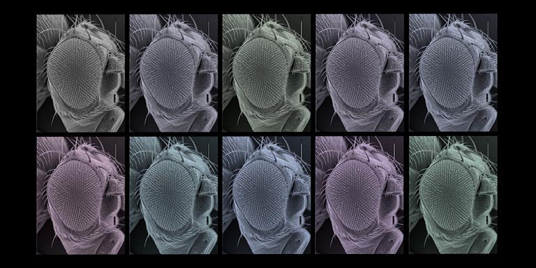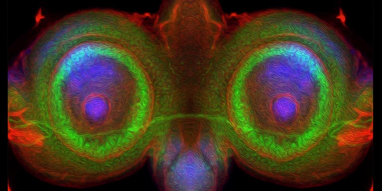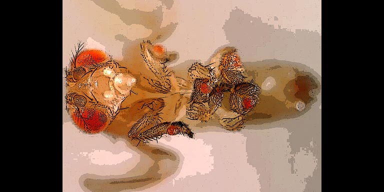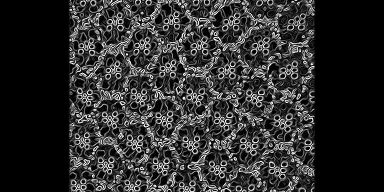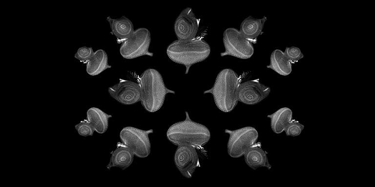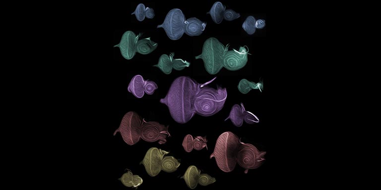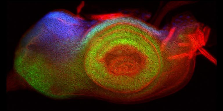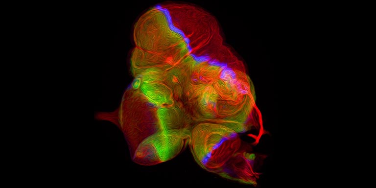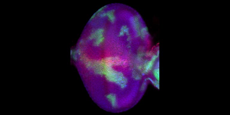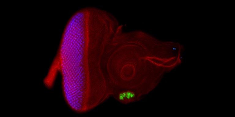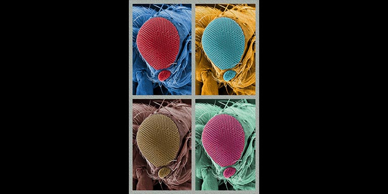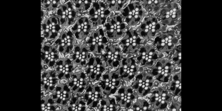

Justin Kumar is a professor of biology and an accidental artist.
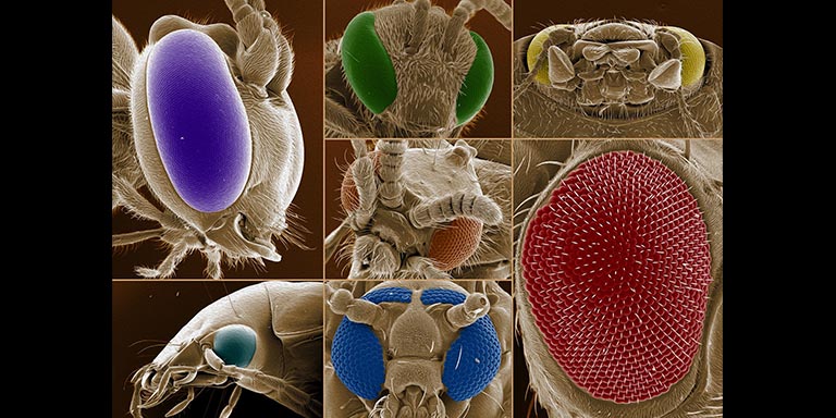
He has been researching flies, specifically Drosophila melanogaster, or the common fruit fly, since his undergraduate years. He didn’t realize then that his research photographs would take on an artistic life of their own. But the art speaks for itself.
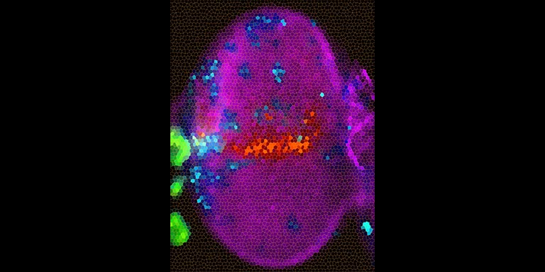
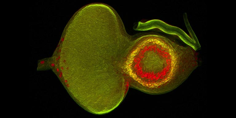
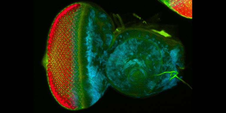
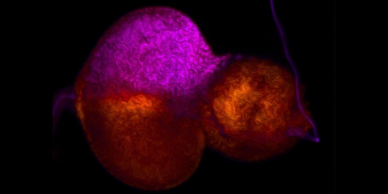
In much of his current research, Professor Kumar’s lab has been researching cell specification – how some cells become an eye instead of, say, a liver.